|
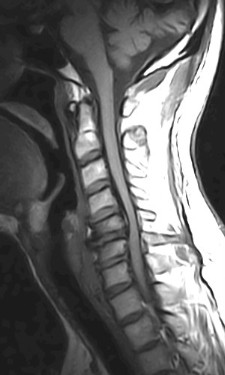
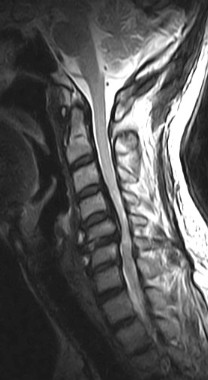
There is evidence of a markedly
thickened/ossified posterior longitudinal ligament over the C3
to the C7 vertebral levels with resultant canal stenosis and
cord compression. The cord shows a hyperintense signal on the
T2W images and is also decreased in caliber (edema/ischemia/gliosis
- myelomalacia).
OPLL may be classified into four types on the
sagittal MR images.
-
Continuous - extending over several
vertebral bodies.
-
Segmental - multiple separate retrovertebral
lesions
-
Mixed - a combination of continuous and
segmental
-
Circumscribed - confined to the retrodiscal
space
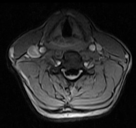
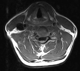
OPLL may be classified into different
morphological types on the axial CT or MR images.
Continuous type is usually thicker, may contain
bone marrow and is most frequently associated with severe cord
compression. Detection is dependent upon the morphology of the
process, presence or absence of bone marrow or calcium in the
ligament or by it's effect upon the ventral subarachnoid space,
dura and spinal cord. Hyperintense signal on the T1W images may
represent fatty marrow. The hypertrophied ligament is
hypointense. Intense enhancement within the ligament may be
seen. T2W images help to assess the cord (myelopathy - due to
direct compression on the spinal cord and anterior spinal
artery).
|